| |
X-ray emission spectroscopy (XES) measures the energy dependence of the emitted X-ray from the decay of the excited state caused by X-ray absorption.
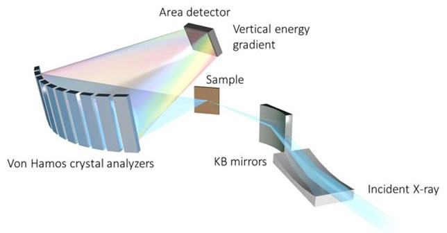
The multilayer monochromator at 25-ID will allow sub-second integration time for mapping. The graphic at right shows how KB mirrors can be used to focus photons onto the sample, while a miniature Von Hamos spectrometer directs flourescence to an area detector (1).
|
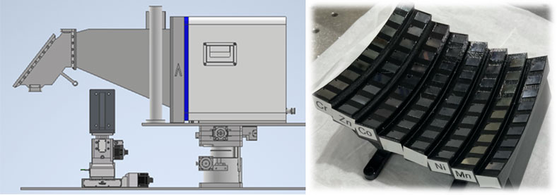
(1) Von Hamos spectrometer design
(2) Next generation multi-element spectrometer Sun, C.-J.; Solovyev, M. A.; Heald, S. et.al. Advanced X-Ray Emission Spectrometers. US Patent 2023/0288352 A1, 2023. |
Data Collection Examples | |
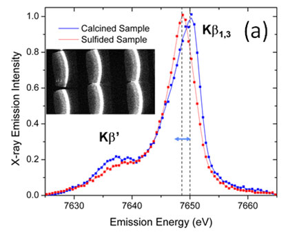
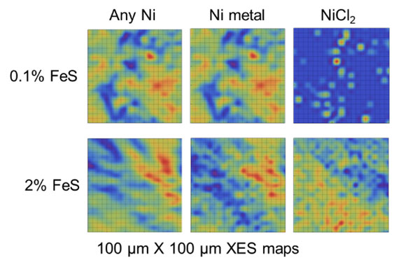
Below are two examples of XES data. On the left is a Co emissions spectra from a catalyst using mini-XS (3). The second shows the data distinguishing Ni and NiCl2 in a battery electrode (4). | |
(3) Co emissions from a catalyst using mini-XS The Kβ1,3 peak shifts for Co with oxygen conpared to sulfur ligands |
(4) Ni and NiCl2 in a battery electrode. Bowden et al, J. Power Sources 247, 517-526 (2014) |
Official websites use .gov
A .gov website belongs to an official government organization in the United States.
Secure .gov websites use HTTPS
A lock (
) or https:// means you’ve safely connected to the .gov website. Share sensitive information only on official, secure websites.