| |
XRF And Confocal Microscopy | |
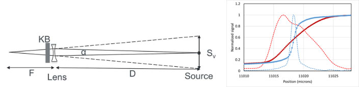
The microprobe beamline at 25-ID-D is optimized for XRF mapping and micro-spectroscopy, including several state-of-the-art fluorescence detectors. Capabilities include chemical mapping with rapid variable focus from 0.5 to 10 microns using unique Be lenses.
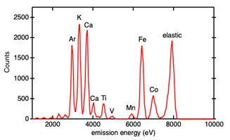
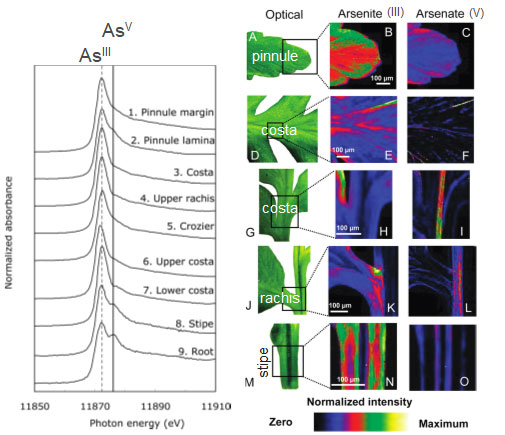
Example XRF spectrum from a sample containing the elements listed. By plotting the signal strength of one element over a 2D sample area, an element specific picture can be produced like those below for As in plant tissue. |
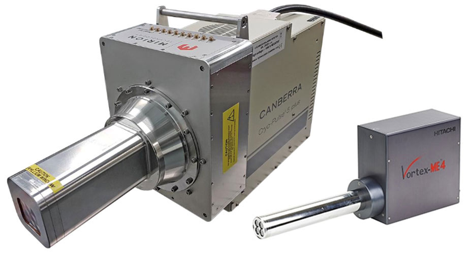
Some of the detectors available for XRF mapping |
Imaging on Resonance
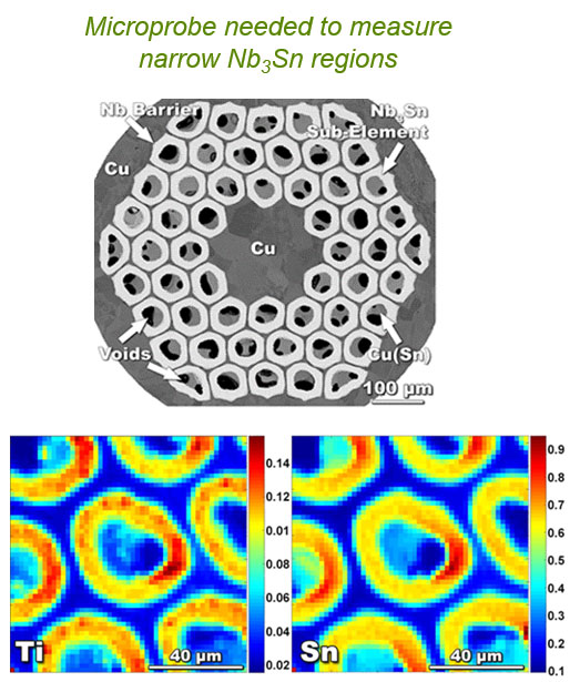
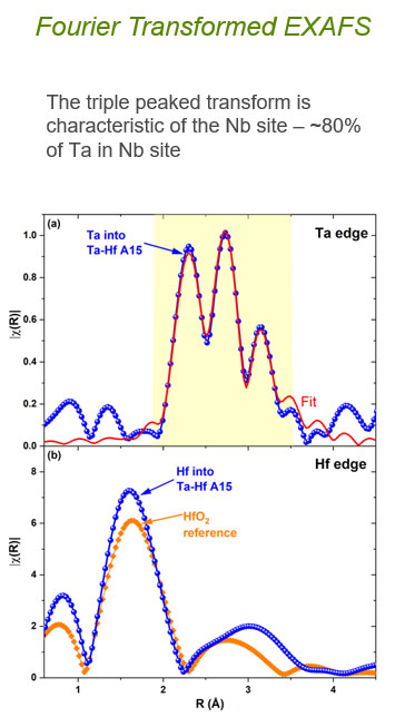
Incident energy held at maximum for an atomic species allows speciation maps. | EXAFS study of Nb3Sn superconductors
Nb3Sn proposed for future accelerator upgrades, but needs improved properties. |
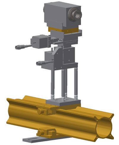
Beryllium Focusing Lenses | |
The beam spot-size can be adjusted quickly and easily through the use of a series of Be lenses contained in a chamber like the one shown. This method has proven to be both reliable and repeatable. | |
| |
Official websites use .gov
A .gov website belongs to an official government organization in the United States.
Secure .gov websites use HTTPS
A lock (
) or https:// means you’ve safely connected to the .gov website. Share sensitive information only on official, secure websites.